


Protein research continues to evolve with advanced technologies, automated systems, and powerful bioinformatics tools. However, some classic laboratory techniques remain essential because of their reliability and analytical depth. One such technique is 2D gel electrophoresis. Even in an era dominated by high-throughput methods, you still benefit greatly from using 2D gels when studying complex protein mixtures.
When you work with biological samples, proteins rarely behave in simple ways. Post-translational modifications, isoforms, degradation products, and subtle expression differences can dramatically influence research outcomes. If you want to detect these variations clearly, you need a method capable of separating proteins with high resolution. This is where 2D gel electrophoresis continues to prove its value.
Understanding the Power of 2D Gel Separation
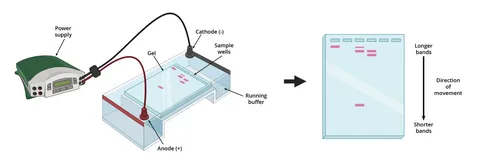
Two-dimensional gel electrophoresis separates proteins based on two independent properties: isoelectric point and molecular weight. In the first dimension, proteins separate through isoelectric focusing. In the second dimension, SDS-PAGE separates them by size.
Because of this dual separation process, you can visualize hundreds or even thousands of proteins within a single gel. Each spot on the gel represents a distinct protein species, making it easier to observe subtle differences that may not appear in simpler techniques.
When you explore detailed separation capabilities, you begin to understand why researchers still rely on high-resolution laboratory methods using 2d Gels for complex protein separation and analysis. These gels reveal patterns that many modern methods may overlook.
Detecting Subtle Protein Variations
Modern protein analysis often focuses on identifying very small biological changes. These changes can include phosphorylation, glycosylation, or minor shifts in protein expression levels.
If you rely only on automated detection systems, some of these differences may remain hidden. 2D gels allow you to visually compare samples and observe protein spot shifts directly. Even a small movement in the gel pattern can signal an important biological modification.
For example, when comparing treated and untreated samples, you might notice a protein spot that shifts slightly due to a post-translational modification. This visual evidence provides an immediate clue that further investigation is needed.
By carefully examining these patterns, you gain insights into cellular mechanisms, disease progression, and biochemical responses.
Ideal for Complex Protein Mixtures
Biological samples rarely contain a single protein. Instead, they consist of thousands of proteins interacting with one another. Analyzing such complexity requires a technique capable of resolving crowded protein mixtures.
2D gels excel in this situation because they spread proteins across a two-dimensional matrix. Instead of stacking proteins in a single line like traditional electrophoresis, they distribute them across the gel surface.
This expanded separation allows you to distinguish proteins that would otherwise overlap. As a result, you can analyze proteins from tissues, cells, bacteria, and biological fluids more effectively.
For researchers working with complicated samples, this level of resolution is critical for generating reliable results.
Visual Protein Mapping for Comparative Studies
Another reason 2D gels remain valuable is their ability to create protein maps. These maps act as fingerprints for biological samples.
When you run multiple gels under controlled conditions, you can compare patterns across different experiments. This helps you identify which proteins appear, disappear, or change intensity between samples.
Such comparisons are extremely useful when studying:
- Disease vs. healthy samples
- Drug treatment responses
- Environmental stress in cells
- Genetic modifications
By comparing protein maps side by side, you quickly detect changes that might indicate new biological pathways or protein interactions.
Supporting Downstream Protein Identification
Although 2D gels provide excellent separation, they also work well with modern identification methods. After separating proteins on a gel, you can excise specific spots and analyze them using mass spectrometry or other analytical tools.
This workflow combines the strengths of classic electrophoresis with advanced molecular analysis. You first isolate proteins visually, then confirm their identities using precise detection technologies.
If you want reliable data from your protein studies, working with an experienced laboratory makes a difference. Many researchers turn to trusted protein analysis expertise available through Kendrick Labs, Inc advanced protein analysis laboratory services to ensure accurate gel separation and interpretation.
Reliable for Long-Term Research Projects
Consistency is essential in scientific research. When you conduct long-term studies, you need techniques that produce reproducible results across multiple experiments.
2D gel electrophoresis has been refined over decades, making it a stable and dependable method. When performed under controlled conditions, gels produce consistent protein patterns that allow accurate comparisons over time.
This reliability is especially useful in pharmaceutical development, biomarker discovery, and clinical research.
Complementing Modern Proteomics
Instead of replacing older methods, modern proteomics often works best when combined with traditional approaches. 2D gels provide an initial visual screening step that highlights important protein changes.
Once you detect interesting patterns, you can apply more specialized techniques to analyze those proteins in detail. This layered approach improves both accuracy and efficiency in protein research.
In other words, 2D gels act as a powerful starting point that guides further investigation.
When You Should Consider Using 2D Gels
You may benefit from using 2D gel analysis if you need to:
- Compare complex protein mixtures
- Detect post-translational modifications
- Visualize protein expression differences
- Map protein profiles across samples
- Prepare proteins for downstream identification
If your project involves subtle protein changes, this technique can provide insights that other methods miss.
For researchers seeking expert guidance or laboratory services, you can always contact us today for professional protein analysis support and consultation to discuss your specific experimental needs.
Conclusion
Despite the rapid growth of automated proteomics technologies, 2D gel electrophoresis continues to play an important role in modern protein analysis. Its ability to separate proteins based on two distinct properties gives you a detailed view of complex biological samples.
When you combine the visual clarity of protein maps with modern identification techniques, you create a powerful analytical workflow. This is why many researchers still rely on 2D gels for detecting subtle protein changes, studying biological complexity, and supporting advanced proteomics research.
If your work requires precise protein separation and reliable comparison between samples, 2D gel analysis remains one of the most valuable tools available.
Frequently Asked Questions
What are 2D gels used for in protein analysis?
2D gels are used to separate proteins based on isoelectric point and molecular weight. This allows researchers to analyze complex protein mixtures and detect subtle changes in protein expression.
Why are 2D gels still important in modern proteomics?
2D gels remain important because they provide high-resolution protein separation and visual protein mapping, helping researchers detect modifications and expression differences.
How many proteins can be detected using 2D gel electrophoresis?
A single 2D gel can separate hundreds to thousands of proteins, depending on the sample complexity and gel conditions.
Can 2D gels work with modern protein identification methods?
Yes. After separation, individual protein spots can be removed from the gel and analyzed using techniques like mass spectrometry for precise identification.
What type of samples work best with 2D gel electrophoresis?
2D gels work well with complex biological samples such as tissue extracts, cell lysates, bacterial proteins, and biological fluids where multiple proteins need to be separated and analyzed.